The Molecular Mechanics of Genetic Duplication
The transmission of biological information from one generation to the next depends entirely on the high-fidelity copying of deoxyribonucleic acid (DNA). This process, known as DNA replication, is a...
The transmission of biological information from one generation to the next depends entirely on the high-fidelity copying of deoxyribonucleic acid (DNA). This process, known as DNA replication, is a masterpiece of molecular engineering, requiring the coordinated action of dozens of specialized proteins to unwind, copy, and proofread the genetic code. Because the structure of the DNA double helix is inherently directional and chemically stable, the cell must employ sophisticated dna replication enzymes to overcome the energetic barriers of synthesis. By examining the steps of dna replication, we reveal a biological system that balances extreme speed with nearly perfect accuracy. This molecular choreography ensures that every time a cell divides, the resulting daughter cells receive a complete and functional set of genetic instructions.
The Semiconservative Nature of Heredity
Before the mechanical details of replication were understood, biologists debated how the double-stranded molecule could be copied without losing its structural integrity. In 1958, Matthew Meselson and Franklin Stahl performed an experiment using heavy ($^{15}N$) and light ($^{14}N$) nitrogen isotopes to track DNA density across generations of Escherichia coli. Their results provided definitive evidence for the semiconservative replication model, which posits that each of the two parental strands serves as a template for a new partner. Consequently, every new DNA molecule is a hybrid, containing one "old" strand and one "new" strand. This mechanism is elegant because it utilizes the inherent complementarity of the base pairs—adenine with thymine and cytosine with guanine—to ensure that the sequence is preserved.
The distinction between the parent strand and the template strand is more than semantic; it is a matter of chemical necessity. The parental double helix is held together by hydrogen bonds between nitrogenous bases, which must be systematically broken to expose the information within. As the strands separate, they act as physical scaffolds upon which the replication machinery assembles. The architectural integrity of the double helix is such that the strands are antiparallel, meaning one runs in the 5' to 3' direction while the other runs 3' to 5'. This orientation creates a significant challenge for the enzymes involved, as DNA synthesis can only proceed in a single chemical direction.
The thermodynamic stability of the B-DNA form—the most common helical structure—presents a barrier to the initiation of replication. The nitrogenous bases are buried within the hydrophobic core of the helix, protected from the aqueous environment of the nucleus or cytoplasm. To access this code, the cell must invest metabolic energy to destabilize the helix at specific locations. This investment is the first critical phase in the steps of dna replication, setting the stage for the massive enzymatic assembly that follows. Without the semiconservative framework, the error rate of replication would likely be too high to support the complexity of multi-cellular life.
Initiation and the Replication Fork
Replication does not begin at random points along the chromosome; instead, it starts at specific sequences known as origins of replication (Ori). In prokaryotes, a single origin is usually sufficient to copy the entire circular genome, but eukaryotic organisms possess thousands of origins spread across their linear chromosomes. This multi-origin strategy is essential for eukaryotes because their genomes are significantly larger, and relying on a single start point would take weeks rather than hours. Specialized initiator proteins recognize these sequences and recruit the machinery necessary to "melt" the DNA, creating a localized bubble where synthesis can commence.
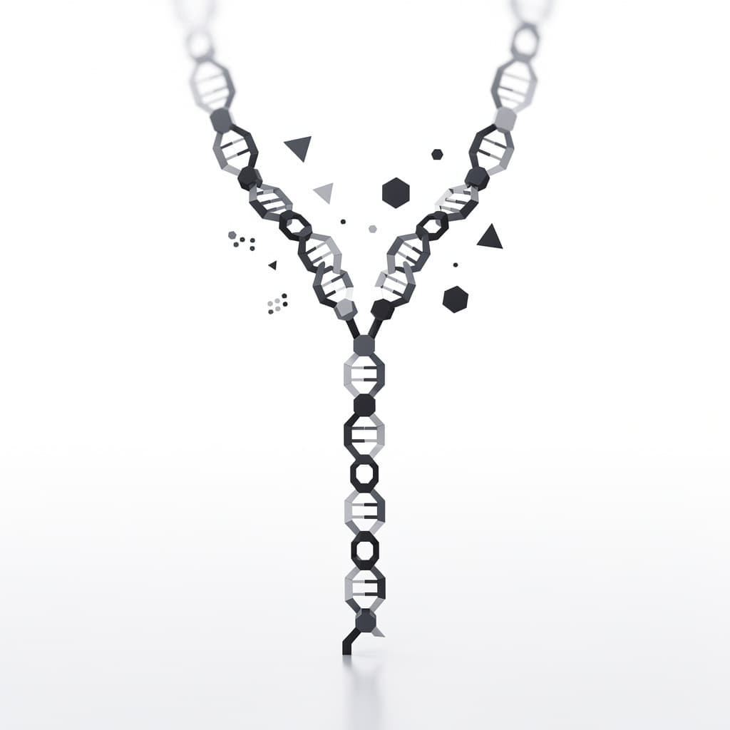
At the heart of the initiation process is DNA helicase, an enzyme that functions like a molecular zipper, consuming ATP to break the hydrogen bonds between base pairs. As helicase unwinds the DNA, it creates a Y-shaped structure known as the replication fork. However, unwinding a tightly coiled helix creates "supercoiling" or torsional strain ahead of the fork, much like twisting a rubber band until it knots. To prevent the DNA from snapping, an enzyme called topoisomerase (or DNA gyrase in bacteria) makes temporary cuts in the phosphate backbone to relieve this tension. This allows the replication fork to move forward at speeds reaching 1,000 nucleotides per second in some species.
Once the two strands are separated, they are highly unstable and prone to re-annealing or forming secondary structures like "hairpins" that would block synthesis. To solve this, single-strand binding proteins (SSBs) coat the exposed DNA, keeping the strands apart and chemically accessible. These proteins do not cover the bases themselves but rather bind to the sugar-phosphate backbone, ensuring the template remains a linear, readable surface. The establishment of the replication fork and the stabilization of the template are the prerequisite steps of dna replication that allow the actual synthesis of new DNA to begin. This phase ensures that the environment is chemically "primed" for the high-speed polymerization that follows.
Primary Enzymes of DNA Synthesis
The primary workhorse of genetic duplication is DNA polymerase, an enzyme that catalyzes the formation of the phosphodiester bond between the 3' carbon of the existing strand and the 5' phosphate of an incoming nucleotide. The dna polymerase function is highly specific; it can only add nucleotides to a pre-existing 3' hydroxyl (-OH) group. This means that DNA polymerase is incapable of starting a new strand from scratch; it can only extend an existing one. This requirement for a "primer" is one of the most curious features of molecular biology, suggesting an evolutionary link to the older "RNA world" where RNA served as the primary genetic material.
To provide this necessary starting point, an enzyme called DNA primase synthesizes a short stretch of RNA, typically 10 to 12 nucleotides long, that is complementary to the DNA template. This RNA primer provides the essential 3'-OH group that DNA polymerase needs to begin its work. Because DNA polymerase is an "instructive" enzyme, it reads the template strand and selects the matching deoxynucleoside triphosphate (dNTP) from the surrounding medium. The energy required for this reaction is provided by the cleavage of two phosphate groups from the incoming dNTP, a process that is thermodynamically favorable and drives the reaction forward. The resulting elongation always occurs in the 5' to 3' direction, a constraint that dictates the entire geometry of the replication fork.
DNA polymerases are also known for their high processivity, meaning they can add thousands of nucleotides without falling off the template. This is often achieved through the use of a "sliding clamp," a ring-shaped protein that tethers the polymerase to the DNA. The precision of dna polymerase function is enhanced by its "palm, fingers, and thumb" domain structure, which allows it to sense the geometry of the base pair being formed. If an incorrect nucleotide is inserted, the geometry is distorted, and the polymerase stalls. This physical check is the first line of defense against mutations, ensuring that the steps of dna replication maintain the fidelity of the genome.
The Leading and Lagging Strand
The antiparallel nature of DNA creates a fundamental problem: as the replication fork opens, one strand runs 3' to 5' toward the fork, while the other runs 5' to 3' toward the fork. Since DNA polymerase can only synthesize in the 5' to 3' direction, the two strands must be handled differently. The leading strand is the one where synthesis moves in the same direction as the replication fork. On this strand, after a single RNA primer is laid down, DNA polymerase can add nucleotides continuously, following the helicase as it unwinds the DNA. This is the most straightforward part of the replication process, requiring minimal coordination once the fork is established.
The lagging strand, however, presents a significant mechanical challenge because its synthesis must move away from the replication fork to maintain the 5' to 3' polarity. To solve this, the cell uses a discontinuous method where the DNA is synthesized in short bursts called Okazaki fragments. As the fork opens further, primase creates a new RNA primer, and DNA polymerase extends it until it hits the previously synthesized fragment. This results in a "back-stitching" mechanism where the lagging strand is built in pieces, each roughly 100 to 200 nucleotides long in eukaryotes and 1,000 to 2,000 in prokaryotes. This discontinuous growth is one of the most complex steps of dna replication, requiring constant resetting of the enzymatic machinery.
The coordination between leading and lagging strand synthesis is managed by the replisome, a massive multi-protein complex that links the two polymerases together. Recent research suggests a "trombone model," where the lagging strand template loops back through the replisome, allowing both polymerases to move in the same physical direction even though they are moving in opposite chemical directions. This synchronization ensures that the replication fork moves as a single unit, preventing the formation of large gaps of vulnerable single-stranded DNA. The interplay between the leading and lagging strand highlights the incredible spatial organization required to overcome the chemical limitations of the DNA molecule.
Finalization and Error Correction
Once the bulk of the DNA has been synthesized, the replication process enters a finalization phase to ensure the new strands are continuous and error-free. The RNA primers used to initiate synthesis must be removed, as they are chemically distinct from DNA and would interfere with the stability of the helix. In many organisms, DNA Polymerase I performs this task; it uses its 5' to 3' exonuclease activity to chew away the RNA and its polymerase activity to replace it with the correct DNA nucleotides. This leaves behind a "nick" in the sugar-phosphate backbone—a missing covalent bond between adjacent fragments.
The enzyme DNA ligase is then recruited to seal these nicks. Ligase uses ATP to catalyze the formation of the final phosphodiester bond, effectively "gluing" the Okazaki fragments on the lagging strand into a single, continuous molecule. While synthesis is occurring, DNA polymerase also performs proofreading through 3' to 5' exonuclease activity. If an incorrect base is added, the enzyme can "back up," remove the mismatched nucleotide, and replace it with the correct one. This self-correcting mechanism reduces the error rate of replication from one in $10^5$ to approximately one in $10^7$ nucleotides, a remarkable level of precision.
A unique problem arises at the ends of linear eukaryotic chromosomes, known as the "end-replication problem." Because the lagging strand requires an RNA primer, there is no way to replace the very last primer at the tip of the chromosome, leading to a slight shortening of the DNA with every round of division. To combat this, cells utilize telomeres—repetitive, non-coding sequences that act as protective caps. In certain cells, such as stem cells and germ cells, an enzyme called telomerase adds these repeats back, preventing the loss of vital genetic information. This final step is crucial for maintaining the "Hayflick limit" and governing the lifespan of cellular lineages.
Cellular Context and Regulation
DNA replication does not happen in isolation; it is strictly regulated to occur only once per cell cycle during the S phase (Synthesis phase). This timing is controlled by a suite of proteins called cyclins and cyclin-dependent kinases (CDKs), which ensure that the replication machinery is assembled only when the cell has sufficient nutrients and lacks DNA damage. If replication were to occur more than once, the cell would end up with extra copies of its genome, a condition known as polyploidy that is often fatal or characteristic of cancerous transformations. The checkpoints governing the steps of dna replication are some of the most highly conserved features in all of biology.
There are notable differences between prokaryotic and eukaryotic systems regarding the speed and scale of replication. Prokaryotic replication is generally faster, with E. coli capable of duplicating its 4.6 million base pair genome in about 40 minutes. Eukaryotes, despite having multiple origins, replicate more slowly because the DNA is tightly wrapped around histone proteins to form chromatin. The replication machinery must navigate this "chromatin landscape," temporarily displacing histones and then reassembling them onto the two new daughter helices. This added layer of complexity involves chaperone proteins that ensure the epigenetic markings—the "memory" of the cell's state—are also inherited.
The metabolic constraints on replication speed are also significant. A single human cell must synthesize approximately 6 billion base pairs of DNA during each S phase. This requires a massive pool of deoxynucleotide triphosphates, the building blocks of DNA. If the supply of any one nucleotide (A, T, C, or G) is depleted, the replication fork may stall, leading to replication stress. Stalled forks are dangerous because they are prone to breaking, which can cause chromosomal translocations or deletions. Cells have evolved "stress response" pathways to protect stalled forks and restart them once nucleotide levels are restored, highlighting the metabolic integration of the replication process.
Applications in Modern Biotechnology
Our understanding of the steps of dna replication has revolutionized medicine and biotechnology, most notably through the development of the Polymerase Chain Reaction (PCR). Developed by Kary Mullis in 1983, PCR mimics the natural replication process in a test tube to amplify specific DNA sequences millions of times. By using a heat-stable DNA polymerase (Taq polymerase) and synthetic DNA primers, scientists can cycle through temperatures that denature the DNA, allow primers to bind, and extend the new strands. PCR is now a cornerstone of forensic science, medical diagnostics, and genetic research, allowing for the detection of tiny amounts of viral or human DNA.
In addition to PCR, the principles of replication are used in synthetic DNA synthesis and modern sequencing technologies. Sanger sequencing, for example, utilizes "chain-terminating" nucleotides that stop DNA polymerase at specific points, allowing researchers to read the sequence of the template. Furthermore, the pharmaceutical industry targets the replication machinery to treat diseases. Many chemotherapy drugs are designed to interfere with DNA replication in rapidly dividing cancer cells. Some drugs mimic nucleotides to cause premature chain termination, while others inhibit topoisomerase, causing the DNA to become so tangled that the cancer cell undergoes programmed cell death (apoptosis).
Finally, the study of replication enzymes has led to the development of "liquid biopsies" and personalized medicine. By understanding how certain mutations affect dna polymerase function, doctors can predict how a patient might respond to specific treatments. Synthetic biology also seeks to create "orthogonal" replication systems—engineered enzymes and templates that can operate independently of the cell's natural machinery. These advances show that the molecular mechanics of genetic duplication are not just a subject of academic interest but a powerful tool for manipulating the building blocks of life itself. As we continue to refine our knowledge of the replication fork, we open new doors to curing genetic disorders and engineering novel biological functions.
References
- Watson, J. D., & Crick, F. H. C., "Molecular Structure of Nucleic Acids: A Structure for Deoxyribose Nucleic Acid", Nature, 1953.
- Meselson, M., & Stahl, F. W., "The Replication of DNA in Escherichia coli", Proceedings of the National Academy of Sciences, 1958.
- Alberts, B., et al., "Molecular Biology of the Cell", 6th Edition, Garland Science, 2014.
- Kornberg, A., "DNA Synthesis", W. H. Freeman and Co., 1974.
- Okazaki, R., et al., "Mechanism of DNA Chain Growth. I. Possible Discontinuity and Unusual Secondary Structure of Newly Synthesized Chains", PNAS, 1968.
Recommended Readings
- The Double Helix by James D. Watson — A first-hand account of the discovery of the DNA structure that provides context for how replication was first conceptualized.
- Lehninger Principles of Biochemistry by David L. Nelson and Michael M. Cox — A foundational textbook that explains the energetic and chemical mechanisms of polymerase activity in exquisite detail.
- DNA Replication by Arthur Kornberg and Tania Baker — Often considered the "bible" of replication studies, this book dives deep into the enzymology and historical experiments of the field.
- The Eighth Day of Creation by Horace Freeland Judson — A masterful historical narrative of the molecular biology revolution that describes the intellectual struggle to understand genetic duplication.